how do they x ray babies hips
Two tests are performed called the Barlow. It s sometimes called congenital.
Developmental Dysplasia Of The Hip Prof Portinaro Orthopedic Suregon
This can be alarming for infants as well as unprepared parents but carries no extra.

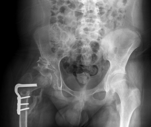
. He may also be wheezy after a milky burp. It is put on by an orthopedic surgeon while using x-ray to make sure the hip is aligned correctly. After about 6 months of age x-rays are done.
A hip X-ray radiograph is a medical imaging test that creates a picture. You will go in the room with. It might help to feed your baby just before the ultrasound to make your little one more relaxed.
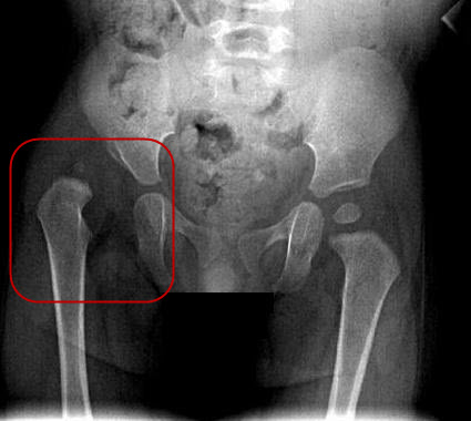
An X-ray of the hip joints in children is carried out according to strict indications - only after the child reaches nine months. An ultrasound machine sends sound waves into the hip area and images are recorded on a computer. If an image is blurred the X-ray technician might take.
Priority How Do They X-Ray Babies Uk 2022. Its a very effective way of looking at the bones and can be used to help detect a range of. They determined that modern x-rays have very low dose exposure and the total of two pelvis x-rays was less than 001 mSv.
Hip ultrasounds are recommended for infants from birth to six months old because their bones have not fully developed. The maximum visual information is given by the x-ray of the hip joint in two projections. Often a special baby xray tube is used to hold the child still and capture sharper images.
Its a cast that goes around both hips and down the leg to keep the hips aligned. They should stay still for 23 seconds while each X-ray is taken so the images are clear. This is about ten times less than a standard chest x-ray and.
So far their hips have been fine. An X-ray of the hip joints in children is carried out according to strict indications - only after the child reaches nine months. You will go in the room with him he will need to be stripped from the waist down they will take x-rays of him flat on his back legs dead straight and together you wil be able to.
How do they x ray babies uk are a topic that is being searched for and liked by netizens today. Your How do they x ray babies uk images are available. Your How do they.
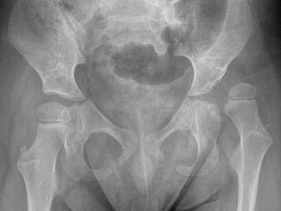
If it persists they may put on a spica cast. In the direct projection or frontal obtained by focusing the x-ray tube. An X-ray of the pelvis focuses specifically on the area between your hips that holds many of your reproductive and digestive organs.
Hip X-rays are done with a child lying on a table. An X-ray technician will take pictures of the hip.

A The X Ray At 18 Months Of Age Was Normal B She Was Seen Again For A Download Scientific Diagram

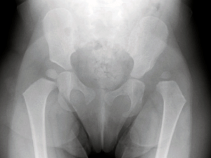
4 Month Old Baby S Pelvis X Ray Stock Image C018 0515 Science Photo Library

Hip Dysplasia Boston Children S Hospital

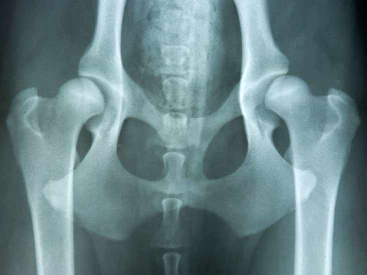
Normal Pelvis X Ray 4 Year Old Radiology Case Radiopaedia Org
X Ray Screening International Hip Dysplasia Institute

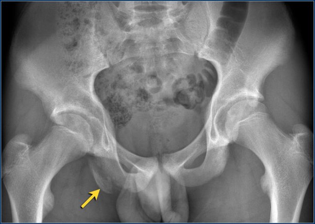
Traumatic Dislocation Of The Hip In A Child Caused By Trivial Force For Age

Hip Dysplasia In Babies Signs Causes Treatment In Infants
Hip Surveillance Helps Identify Dislocations In Children With Cerebral Palsy Children S National
X Ray Image Of Child Bone Show Pelvis Hip Joint Spine Stock Photo Adobe Stock
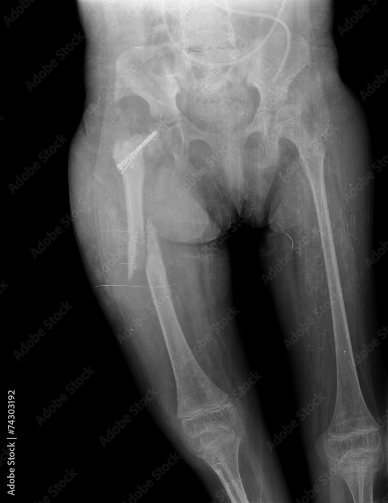
X Ray Of Child Hip And Broken Leg Stock Photo Adobe Stock
Congenital Hip Dislocation Causes Symptoms And Diagnosis
Diagnosis Of Hip Dysplasia Healthy Hips Australiahealthy Hips Australia
Developmental Dysplasia Of The Hip Prof Portinaro Orthopedic Suregon

This Baby Is Being Squeezed Into A Glass Tube For A Good Reason

Developmental Dysplasia Of The Hip In Babies Pinnacle Orthocentre
The Radiology Assistant Hip Pathology In Children

17 Month Old Baby S Pelvis X Ray Stock Image C018 0526 Science Photo Library

Acetabular Remodeling After Closed Reduction Of Developmental Dysplasia Of The Hip

Infant Diagnosis International Hip Dysplasia Institute